Embed Size (px)
DESCRIPTION
nanti kalo berdarah yausdah
Citation preview

Larsson et al. BMC Research Notes 2014, 7:236http://www.biomedcentral.com/1756-0500/7/236
SHORT REPORT Open Access
Chronic non-bloody diarrhoea: a prospectivestudy in Malmö, Sweden, with focus onmicroscopic colitisJohanna K Larsson1, Klas Sjöberg1*, Lina Vigren2, Cecilia Benoni1, Ervin Toth1 and Martin Olesen3
Abstract
Background: Chronic non-bloody diarrhoea affects up to 5% of the population. Microscopic colitis is one of themost common causes, encompassing the subtypes collagenous colitis and lymphocytic colitis. The diagnosis ofmicroscopic colitis is made by histological examination of colonic mucosal biopsy specimens. The aim of thisinvestigation was to determine whether laboratory parameters or questions about disease history or concomitantdisease could be helpful in discriminating patients with MC from those with a histologically normal colonic mucosa.
Findings: Patients admitted for colonoscopy because of chronic non-bloody diarrhoea (>2 loose stoolsfor >3 weeks) at endoscopy units in Malmö during 2007 and 2009, were enrolled. A total number of 78 patientswere included (60 women, 18 men, median age 59, IQR 45–69 years). Out of these 78, 15 patients (19%) hadmicroscopic colitis (CC; n = 10, LC; n = 5). MC was especially prevalent in patients above the age of 50 (25%). Nodifferences were found between those with normal histology and MC in laboratory analyses (inflammatory and liverparameters). Neither were differences shown in questions regarding symptoms, environmental factors orconcomitant diseases except for an association with celiac disease (p = 0.019) and a trend maybe indicating aninverse association with appendectomy (p = 0.057).
Conclusions: Microscopic colitis is associated with female gender, celiac disease and consumption of NSAIDs.Trends were observed indicating that age above 50 years, acute onset and absence of appendectomy may beassociated with MC. No associations were observed with other symptoms, calprotectin levels or liver parameters.
Keywords: Collagenous colitis, Diarrhoea, Inflammatory bowel disease, Lymphocytic colitis, Microscopic colitis
FindingsBackgroundChronic non-bloody diarrhoea (CNBD) is common inWestern populations. A prevalence of 5% in the back-ground population and 15% among elderly has been sug-gested [1]. CNBD is usually defined as more than twoloose stools per day for more than four weeks [2]. Theunderlying disease behind CNBD is not always obviousand can be difficult to diagnose. Besides extra-intestinalcauses, several gastrointestinal conditions may be contem-plated, such as microscopic colitis (MC), IBS-D (irritablebowel disease with predominantly diarrhoea) or celiac dis-ease (CeD). In patients with other complicating diseases
* Correspondence: [email protected] of Clinical Sciences, Department of Gastroenterology andNutrition, Skåne University Hospital, Lund University, Malmö, SwedenFull list of author information is available at the end of the article
© 2014 Larsson et al.; licensee BioMed CentralCommons Attribution License (http://creativecreproduction in any medium, provided the orDedication waiver (http://creativecommons.orunless otherwise stated.
secondary diarrhoea may occur, such as bacterial over-growth in diabetes or bile acid diarrhoea in Crohn’sdisease or as a consequence after cholecystectomy or ileo-cecal resection [3].Consequently, even though CNBD may have many
causes, MC is important to contemplate since it is diag-nosed in 5-20% according to some investigations [4-9].Histological characteristics of MC are well defined [10].The condition can be divided into two subgroups; col-lagenous colitis (CC) and lymphocytic colitis (LC). Col-lagenous colitis was first described as a case report byCG Lindström in 1976 [11] and LC by Lazenby et al. in1989 [12]. In MC a macroscopically normal or near nor-mal mucosa is observed [10]. Weight loss and abdominalpain may occur [13,14]. Since it is possible to verify sus-pected MC first after examination of biopsy specimens
Ltd. This is an Open Access article distributed under the terms of the Creativeommons.org/licenses/by/4.0), which permits unrestricted use, distribution, andiginal work is properly credited. The Creative Commons Public Domaing/publicdomain/zero/1.0/) applies to the data made available in this article,

Larsson et al. BMC Research Notes 2014, 7:236 Page 2 of 7http://www.biomedcentral.com/1756-0500/7/236
any discriminating information in the patient history orlaboratory parameters would be helpful.Occasional previous investigations on patients with ac-
tive MC have noticed a slight elevation in the calprotec-tin levels, indicating an inflammatory response [15,16].Even if this test theoretically could be used for diagnos-tic purposes it is somewhat complicated and if bloodsamples could be used instead it would probably be pre-ferred by the majority of the patients. In view of thepresence of this low grade inflammation an increase insedimentation rate, CRP, number of white blood cells oraberrations in the differential count could be contem-plated. However, limited data are available regarding la-boratory and clinical characteristics in MC. In contrastto classic inflammatory bowel disease (IBD), where dis-turbed liver function may occur, no information is, tothe best of our knowledge, available in MC.
Research hypothesisSince MC is manifested through inflammatory activity incolon we imagined that this inflammatory activity couldbe detected in some way in blood and fecal samples aswell. We also imagined that the symptoms, environmen-tal factors, frequency of concomitant diseases (especiallydiseases considered as autoimmune) as well as charac-teristics of disease onset would differ from patients withCNBD but with histologically normal colonic mucosa.
Methods usedPatients admitted for colonoscopy at out-patient clinics(either to private Endoscopy Units in Malmö or to theEndoscopy unit at the Dept of Gastroenterology andNutrition at Skåne University Hospital, Malmö) becauseof CNBD from January 2007 until June 2009 were in-vited to this investigation. CNDB was defined as morethan two loose stools per day for more than three weeks,where blood not had been detected. Those willing toparticipate were asked to answer a questionnaire abouttheir past and present medical history and also left bloodand fecal samples.A self administrated questionnaire about chronic diar-
rhoea onset (acute or slow), stool frequency (“high” ifmore than 10 stools/day), stools at night and consistencywas completed by the patients. They were also askedquestions about recent journeys abroad (i.e. a journeyabroad within a month before onset of symptoms) andabout “surrounding cases of diarrhoea” (if anyone in theimmediate surroundings had had onset of diarrhoea atthe same time as the patient). A list of different potentialco-factors was also penetrated, such as smoking habits,previous surgery (cholecystectomy or appendectomy),and presence of celiac disease, thyroid disease, rheuma-toid arthritis or diabetes mellitus. Medical treatment wasalso taken into consideration with questions concerning
ongoing drug treatment. Three to four years later the pa-tients were asked whether they had recovered or not (totalrecovery or a reduction of symptoms three to four yearsafter colonoscopy compared to before colonoscopy).Blood and fecal samples were collected prior to colonos-
copy. C-reactive protein (CRP), sedimentation rate, haemo-globin, white blood cells with differential count, platelets,creatinine, aspartate transaminase (AST), alanine trans-aminase (ALT), alkaline phosphatase (ALP), bilirubin aswell as fecal calprotectin were analysed at the Departmentof Clinical Chemistry, Skåne University Hospital Malmö,according to standardized procedures. Calprotectin wasanalyzed according to the procedure described by Tønet al. [17].During colonoscopy mucosal biopsy specimen were
obtained from distal ileum, caecum, ascending colon,right flexure, transverse colon, left flexure, descendingcolon, sigmoid colon as well as rectum with at least twobiopsies from each location.The histopathological criteria used for CC were:
� A thickened subepithelial collagen layer ≥ 10micrometers (μm)
� A chronic inflammatory infiltrate in the laminapropria
� Epithelial damage such as flattening and detachment
The histopathological criteria used for LC were:
� Intraepithelial lymphocytes (IEL) ≥ 20 per 100surface epithelial cells
� A subepithelial collagen layer < 10 μm� A chronic inflammatory infiltrate in the lamina propria� Epithelial damage such as flattening and detachment
The specimens were evaluated by a gastrointestinalpathologist with a special interest in MC (M Olesen). Thesubepithelial collagen layer was measured with an ocularmicrometer in a well orientated part of a mucosal section.Measurement of the collagen layer as well as counting ofIEL was performed in haematoxylin-eosin. Whenever con-sidered necessary special stainings for collagen fibres(Masson’s trichrome or van Gieson) or reticulin fibres(Sirius red) were carried out. Whenever considered neededsections were immunohistochemically stained with anti-bodies against T-lymphocytes (CD3) [10]. Diagnoses ofulcerative colitis or Crohn’s disease were set according toestablished criteria previously described in detail [18].The investigation was approved by the local Commit-
tee of Research Ethics at Lund University (LU: 276/2006). The patients were included first after written in-formed consent.For analyses of differences between patients with MC and
those with a normal colonoscopy based on macroscopic
Larsson et al. BMC Research Notes 2014, 7:236 Page 3 of 7http://www.biomedcentral.com/1756-0500/7/236
and microscopic findings (i.e. patients with classic IBDwere excluded) Kruskall-Wallis’ non-parametric test wasused. Chi2 analysis was used to compare frequencies ofMC in different age groups. A p-value below 0.05 wasconsidered significant.
ResultsDuring the study period 397 patients were admitted forcolonoscopy with CNBD. Of these 176 did not answer(mean age 47 years, 108 females (61%)), 110 actively de-nied participation (mean age 58 years, 80 females (73%))and 33 cancelled their colonoscopy (mean age 54 years,
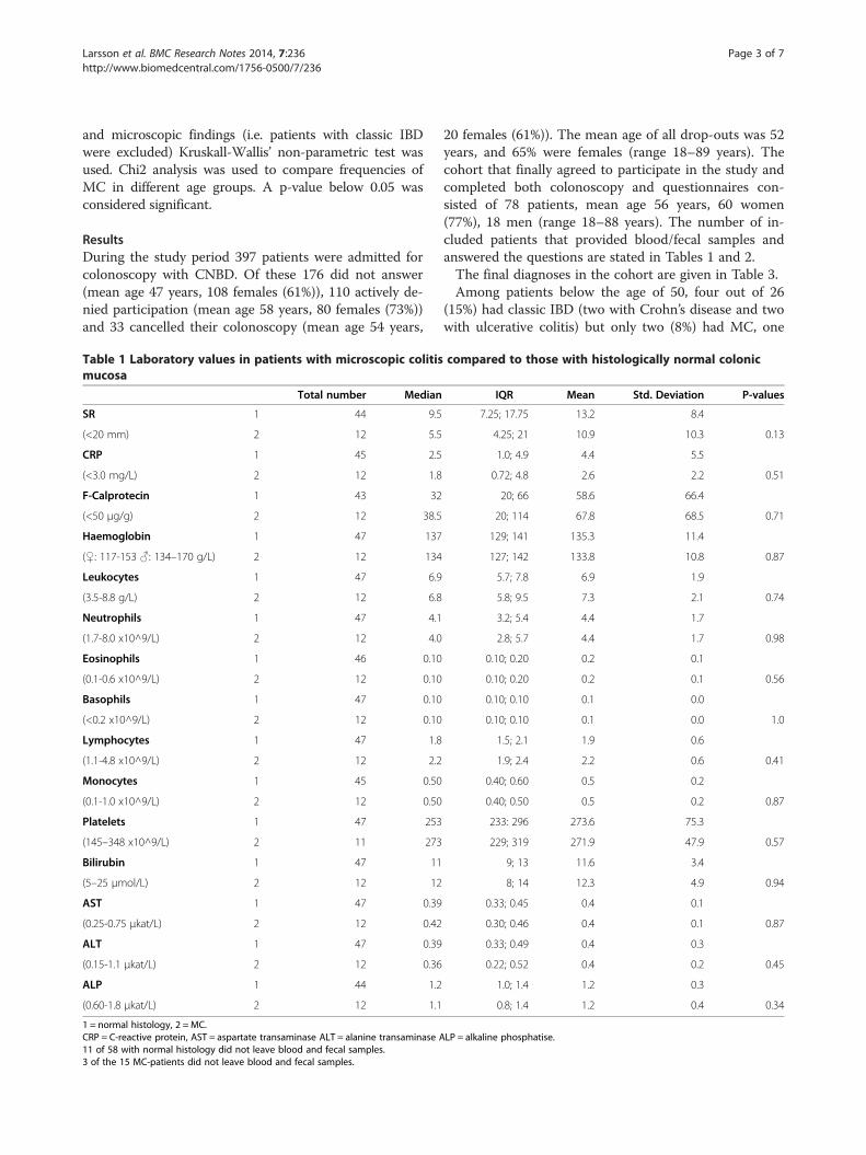
Table 1 Laboratory values in patients with microscopic colitismucosa
Total number Median
SR 1 44 9.5
(<20 mm) 2 12 5.5
CRP 1 45 2.5
(<3.0 mg/L) 2 12 1.8
F-Calprotecin 1 43 32
(<50 μg/g) 2 12 38.5
Haemoglobin 1 47 137
(♀: 117-153 ♂: 134–170 g/L) 2 12 134
Leukocytes 1 47 6.9
(3.5-8.8 g/L) 2 12 6.8
Neutrophils 1 47 4.1
(1.7-8.0 x10^9/L) 2 12 4.0
Eosinophils 1 46 0.10
(0.1-0.6 x10^9/L) 2 12 0.10
Basophils 1 47 0.10
(<0.2 x10^9/L) 2 12 0.10
Lymphocytes 1 47 1.8
(1.1-4.8 x10^9/L) 2 12 2.2
Monocytes 1 45 0.50
(0.1-1.0 x10^9/L) 2 12 0.50
Platelets 1 47 253
(145–348 x10^9/L) 2 11 273
Bilirubin 1 47 11
(5–25 μmol/L) 2 12 12
AST 1 47 0.39
(0.25-0.75 μkat/L) 2 12 0.42
ALT 1 47 0.39
(0.15-1.1 μkat/L) 2 12 0.36
ALP 1 44 1.2
(0.60-1.8 μkat/L) 2 12 1.1
1 = normal histology, 2 = MC.CRP = C-reactive protein, AST = aspartate transaminase ALT = alanine transaminase A11 of 58 with normal histology did not leave blood and fecal samples.3 of the 15 MC-patients did not leave blood and fecal samples.
20 females (61%)). The mean age of all drop-outs was 52years, and 65% were females (range 18–89 years). Thecohort that finally agreed to participate in the study andcompleted both colonoscopy and questionnaires con-sisted of 78 patients, mean age 56 years, 60 women(77%), 18 men (range 18–88 years). The number of in-cluded patients that provided blood/fecal samples andanswered the questions are stated in Tables 1 and 2.The final diagnoses in the cohort are given in Table 3.Among patients below the age of 50, four out of 26
(15%) had classic IBD (two with Crohn’s disease and twowith ulcerative colitis) but only two (8%) had MC, one
compared to those with histologically normal colonic
IQR Mean Std. Deviation P-values
7.25; 17.75 13.2 8.4
4.25; 21 10.9 10.3 0.13
1.0; 4.9 4.4 5.5
0.72; 4.8 2.6 2.2 0.51
20; 66 58.6 66.4
20; 114 67.8 68.5 0.71
129; 141 135.3 11.4
127; 142 133.8 10.8 0.87
5.7; 7.8 6.9 1.9
5.8; 9.5 7.3 2.1 0.74
3.2; 5.4 4.4 1.7
2.8; 5.7 4.4 1.7 0.98
0.10; 0.20 0.2 0.1
0.10; 0.20 0.2 0.1 0.56
0.10; 0.10 0.1 0.0
0.10; 0.10 0.1 0.0 1.0
1.5; 2.1 1.9 0.6
1.9; 2.4 2.2 0.6 0.41
0.40; 0.60 0.5 0.2
0.40; 0.50 0.5 0.2 0.87
233: 296 273.6 75.3
229; 319 271.9 47.9 0.57
9; 13 11.6 3.4
8; 14 12.3 4.9 0.94
0.33; 0.45 0.4 0.1
0.30; 0.46 0.4 0.1 0.87
0.33; 0.49 0.4 0.3
0.22; 0.52 0.4 0.2 0.45
1.0; 1.4 1.2 0.3
0.8; 1.4 1.2 0.4 0.34
LP = alkaline phosphatise.
Larsson et al. BMC Research Notes 2014, 7:236 Page 4 of 7http://www.biomedcentral.com/1756-0500/7/236
with CC and one with LC. See Table 3 where all diagno-ses in the population have been listed. In contrast to thisfinding, in patients 50 years old or older only one out of52 had Crohn’s disease (2%) but as many as 13 (25%)had MC; nine CC and four LC (p = 0.067). The age andsex distributions in patients with normal histology andCC and LC, respectively, are depicted in Table 4. As canbe seen in this Table the patients with MC were allwomen compared to 41 out of 58 in those with normalhistology (p = 0.017).Patients with MC (n = 15) were compared with pa-
tients with normal histology (where also patients with
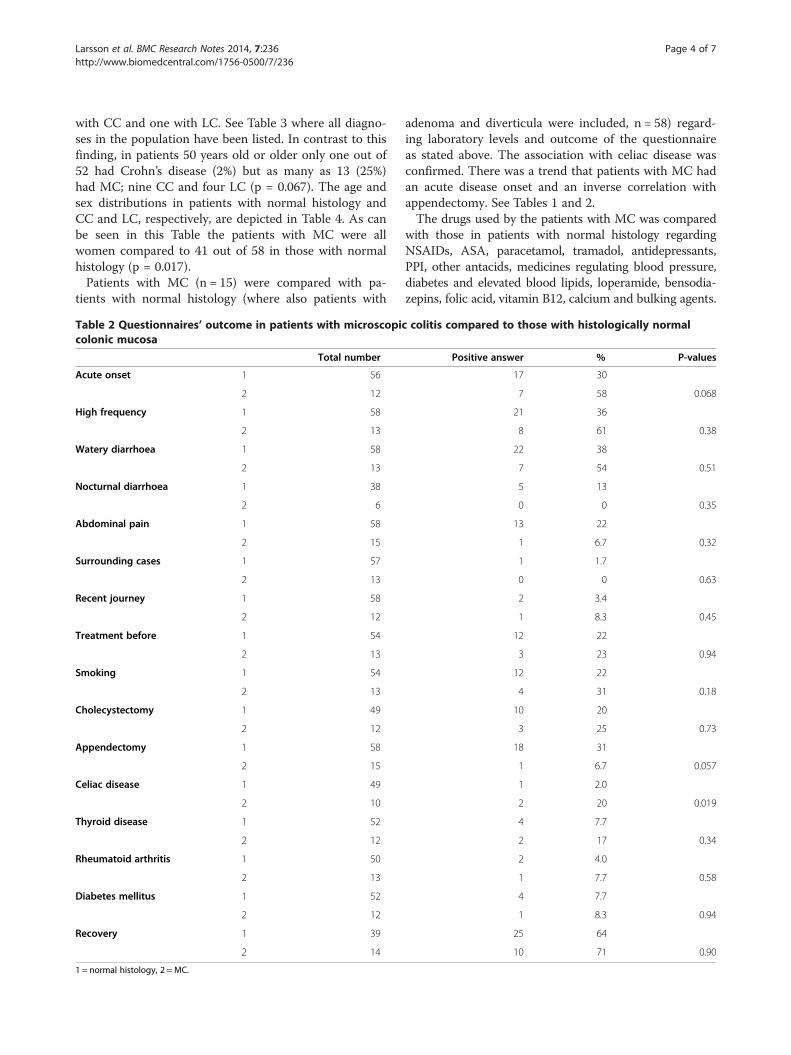
Table 2 Questionnaires’ outcome in patients with microscopiccolonic mucosa
Total number
Acute onset 1 56
2 12
High frequency 1 58
2 13
Watery diarrhoea 1 58
2 13
Nocturnal diarrhoea 1 38
2 6
Abdominal pain 1 58
2 15
Surrounding cases 1 57
2 13
Recent journey 1 58
2 12
Treatment before 1 54
2 13
Smoking 1 54
2 13
Cholecystectomy 1 49
2 12
Appendectomy 1 58
2 15
Celiac disease 1 49
2 10
Thyroid disease 1 52
2 12
Rheumatoid arthritis 1 50
2 13
Diabetes mellitus 1 52
2 12
Recovery 1 39
2 14
1 = normal histology, 2 = MC.
adenoma and diverticula were included, n = 58) regard-ing laboratory levels and outcome of the questionnaireas stated above. The association with celiac disease wasconfirmed. There was a trend that patients with MC hadan acute disease onset and an inverse correlation withappendectomy. See Tables 1 and 2.The drugs used by the patients with MC was compared
with those in patients with normal histology regardingNSAIDs, ASA, paracetamol, tramadol, antidepressants,PPI, other antacids, medicines regulating blood pressure,diabetes and elevated blood lipids, loperamide, bensodia-zepins, folic acid, vitamin B12, calcium and bulking agents.
colitis compared to those with histologically normal
Positive answer % P-values
17 30
7 58 0.068
21 36
8 61 0.38
22 38
7 54 0.51
5 13
0 0 0.35
13 22
1 6.7 0.32
1 1.7
0 0 0.63
2 3.4
1 8.3 0.45
12 22
3 23 0.94
12 22
4 31 0.18
10 20
3 25 0.73
18 31
1 6.7 0.057
1 2.0
2 20 0.019
4 7.7
2 17 0.34
2 4.0
1 7.7 0.58
4 7.7
1 8.3 0.94
25 64
10 71 0.90
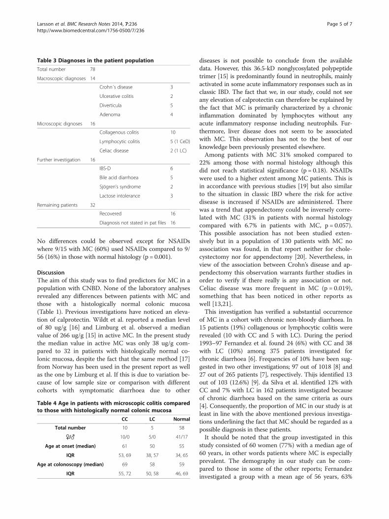
Table 3 Diagnoses in the patient population
Total number 78
Macroscopic diagnoses 14
Crohn´s disease 3
Ulcerative colitis 2
Diverticula 5
Adenoma 4
Microscopic dignoses 16
Collagenous colitis 10
Lymphocytic colitis 5 (1 CeD)
Celiac disease 2 (1 LC)
Further investigation 16
IBS-D 6
Bile acid diarrhoea 5
Sjögren’s syndrome 2
Lactose intolerance 3
Remaining patients 32
Recovered 16
Diagnosis not stated in pat files 16
Larsson et al. BMC Research Notes 2014, 7:236 Page 5 of 7http://www.biomedcentral.com/1756-0500/7/236
No differences could be observed except for NSAIDswhere 9/15 with MC (60%) used NSAIDs compared to 9/56 (16%) in those with normal histology (p = 0.001).
DiscussionThe aim of this study was to find predictors for MC in apopulation with CNBD. None of the laboratory analysesrevealed any differences between patients with MC andthose with a histologically normal colonic mucosa(Table 1). Previous investigations have noticed an eleva-tion of calprotectin. Wildt et al. reported a median levelof 80 ug/g [16] and Limburg et al. observed a medianvalue of 266 ug/g [15] in active MC. In the present studythe median value in active MC was only 38 ug/g com-pared to 32 in patients with histologically normal co-lonic mucosa, despite the fact that the same method [17]from Norway has been used in the present report as wellas the one by Limburg et al. If this is due to variation be-cause of low sample size or comparison with differentcohorts with symptomatic diarrhoea due to other
Table 4 Age in patients with microscopic colitis comparedto those with histologically normal colonic mucosa
CC LC Normal
Total number 10 5 58
♀/♂ 10/0 5/0 41/17
Age at onset (median) 61 50 55
IQR 53, 69 38, 57 34, 65
Age at colonoscopy (median) 69 58 59
IQR 55, 72 50, 58 46, 69
diseases is not possible to conclude from the availabledata. However, this 36.5-kD nonglycosylated polypeptidetrimer [15] is predominantly found in neutrophils, mainlyactivated in some acute inflammatory responses such as inclassic IBD. The fact that we, in our study, could not seeany elevation of calprotectin can therefore be explained bythe fact that MC is primarily characterized by a chronicinflammation dominated by lymphocytes without anyacute inflammatory response including neutrophils. Fur-thermore, liver disease does not seem to be associatedwith MC. This observation has not to the best of ourknowledge been previously presented elsewhere.Among patients with MC 31% smoked compared to
22% among those with normal histology although thisdid not reach statistical significance (p = 0.18). NSAIDswere used to a higher extent among MC patients. This isin accordance with previous studies [19] but also similarto the situation in classic IBD where the risk for activedisease is increased if NSAIDs are administered. Therewas a trend that appendectomy could be inversely corre-lated with MC (31% in patients with normal histologycompared with 6.7% in patients with MC, p = 0.057).This possible association has not been studied exten-sively but in a population of 130 patients with MC noassociation was found, in that report neither for chole-cystectomy nor for appendectomy [20]. Nevertheless, inview of the association between Crohn’s disease and ap-pendectomy this observation warrants further studies inorder to verify if there really is any association or not.Celiac disease was more frequent in MC (p = 0.019),something that has been noticed in other reports aswell [13,21].This investigation has verified a substantial occurrence
of MC in a cohort with chronic non-bloody diarrhoea. In15 patients (19%) collagenous or lymphocytic colitis wererevealed (10 with CC and 5 with LC). During the period1993–97 Fernandez et al. found 24 (6%) with CC and 38with LC (10%) among 375 patients investigated forchronic diarrhoea [6]. Frequencies of 10% have been sug-gested in two other investigations; 97 out of 1018 [8] and27 out of 265 patients [7], respectively. Thijs identified 13out of 103 (12.6%) [9]. da Silva et al. identified 12% withCC and 7% with LC in 162 patients investigated becauseof chronic diarrhoea based on the same criteria as ours[4]. Consequently, the proportion of MC in our study is atleast in line with the above mentioned previous investiga-tions underlining the fact that MC should be regarded as apossible diagnosis in these patients.It should be noted that the group investigated in this
study consisted of 60 women (77%) with a median age of60 years, in other words patients where MC is especiallyprevalent. The demography in our study can be com-pared to those in some of the other reports; Fernandezinvestigated a group with a mean age of 56 years, 63%
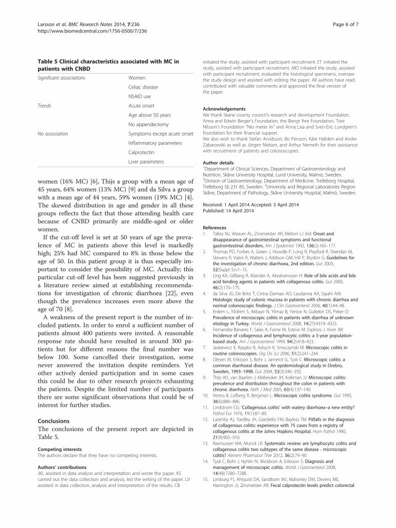
Table 5 Clinical characteristics associated with MC inpatients with CNBD
Significant associations Women
Celiac disease
NSAID use
Trends Acute onset
Age above 50 years
No appendectomy
No association Symptoms except acute onset
Inflammatory parameters
Calprotectin
Liver parameters
Larsson et al. BMC Research Notes 2014, 7:236 Page 6 of 7http://www.biomedcentral.com/1756-0500/7/236
women (16% MC) [6], Thijs a group with a mean age of45 years, 64% women (13% MC) [9] and da Silva a groupwith a mean age of 44 years, 59% women (19% MC) [4].The skewed distribution in age and gender in all thesegroups reflects the fact that those attending health carebecause of CNBD primarily are middle-aged or olderwomen.If the cut-off level is set at 50 years of age the preva-
lence of MC in patients above this level is markedlyhigh; 25% had MC compared to 8% in those below theage of 50. In this patient group it is thus especially im-portant to consider the possibility of MC. Actually; thisparticular cut-off level has been suggested previously ina literature review aimed at establishing recommenda-tions for investigation of chronic diarrhoea [22], eventhough the prevalence increases even more above theage of 70 [8].A weakness of the present report is the number of in-
cluded patients. In order to enrol a sufficient number ofpatients almost 400 patients were invited. A reasonableresponse rate should have resulted in around 300 pa-tients but for different reasons the final number wasbelow 100. Some cancelled their investigation, somenever answered the invitation despite reminders. Yetother actively denied participation and in some casesthis could be due to other research projects exhaustingthe patients. Despite the limited number of participantsthere are some significant observations that could be ofinterest for further studies.
ConclusionsThe conclusions of the present report are depicted inTable 5.
Competing interestsThe authors declare that they have no competing interests.
Authors’ contributionsJKL assisted in data analysis and interpretation and wrote the paper. KScarried out the data collection and analysis, led the writing of the paper. LVassisted in data collection, analysis and interpretation of the results. CB
initiated the study, assisted with participant recruitment. ET initiated thestudy, assisted with participant recruitment. MO initiated the study, assistedwith participant recruitment, evaluated the histological specimens, oversawthe study design and assisted with editing the paper. All authors have read,contributed with valuable comments and approved the final version ofthe paper.
AcknowledgementsWe thank Skane county council’s research and development Foundation,Anna and Edwin Berger’s Foundation, the Bengt Ihre Foundation, ToreNilsson’s Foundation “Nio meter liv” and Anna Lisa and Sven-Eric Lundgren’sfoundation for their financial support.We also wish to thank Stefan Arvidsson, Bo Persson, Kåre Hallden and AndreZabarowski as well as Jörgen Nielsen, and Arthur Nemeth for their assistancewith recruitment of patients and colonoscopies.
Author details1Department of Clinical Sciences, Department of Gastroenterology andNutrition, Skåne University Hospital, Lund University, Malmö, Sweden.2Division of Gastroenterology, Department of Medicine, Trelleborg Hospital,Trelleborg SE-231 85, Sweden. 3University and Regional Laboratories RegionSkåne, Department of Pathology, Skåne University Hospital, Malmö, Sweden.
Received: 1 April 2014 Accepted: 3 April 2014Published: 14 April 2014
References1. Talley NJ, Weaver AL, Zinsmeister AR, Melton LJ 3rd: Onset and
disappearance of gastrointestinal symptoms and functionalgastrointestinal disorders. Am J Epidemiol 1992, 136(2):165–177.
2. Thomas PD, Forbes A, Green J, Howdle P, Long R, Playford R, Sheridan M,Stevens R, Valori R, Walters J, Addison GM, Hill P, Brydon G: Guidelines forthe investigation of chronic diarrhoea, 2nd edition. Gut 2003,52(Suppl 5):v1–15.
3. Ung KA, Gillberg R, Kilander A, Abrahamsson H: Role of bile acids and bileacid binding agents in patients with collagenous colitis. Gut 2000,46(2):170–175.
4. da Silva JG, De Brito T, Cintra Damiao AO, Laudanna AA, Sipahi AM:Histologic study of colonic mucosa in patients with chronic diarrhea andnormal colonoscopic findings. J Clin Gastroenterol 2006, 40(1):44–48.
5. Erdem L, Yildirim S, Akbayir N, Yilmaz B, Yenice N, Gultekin OS, Peker O:Prevalence of microscopic colitis in patients with diarrhea of unknownetiology in Turkey. World J Gastroenterol 2008, 14(27):4319–4323.
6. Fernandez-Banares F, Salas A, Forne M, Esteve M, Espinos J, Viver JM:Incidence of collagenous and lymphocytic colitis: a 5-year population-based study. Am J Gastroenterol 1999, 94(2):418–423.
7. Jaskiewicz K, Rzepko R, Adrych K, Smoczynski M: Microscopic colitis inroutine colonoscopies. Dig Dis Sci 2006, 51(2):241–244.
8. Olesen M, Eriksson S, Bohr J, Jarnerot G, Tysk C: Microscopic colitis: acommon diarrhoeal disease. An epidemiological study in Orebro,Sweden, 1993–1998. Gut 2004, 53(3):346–350.
9. Thijs WJ, van Baarlen J, Kleibeuker JH, Kolkman JJ: Microscopic colitis:prevalence and distribution throughout the colon in patients withchronic diarrhoea. Neth J Med 2005, 63(4):137–140.
10. Veress B, Lofberg R, Bergman L: Microscopic colitis syndrome. Gut 1995,36(6):880–886.
11. Lindstrom CG: ’Collagenous colitis’ with watery diarrhoea–a new entity?Pathol Eur 1976, 11(1):87–89.
12. Lazenby AJ, Yardley JH, Giardiello FM, Bayless TM: Pitfalls in the diagnosisof collagenous colitis: experience with 75 cases from a registry ofcollagenous colitis at the Johns Hopkins Hospital. Hum Pathol 1990,21(9):905–910.
13. Rasmussen MA, Munck LK: Systematic review: are lymphocytic colitis andcollagenous colitis two subtypes of the same disease - microscopiccolitis? Aliment Pharmacol Ther 2012, 36(2):79–90.
14. Tysk C, Bohr J, Nyhlin N, Wickbom A, Eriksson S: Diagnosis andmanagement of microscopic colitis. World J Gastroenterol 2008,14(48):7280–7288.
15. Limburg PJ, Ahlquist DA, Sandborn WJ, Mahoney DW, Devens ME,Harrington JJ, Zinsmeister AR: Fecal calprotectin levels predict colorectal

Larsson et al. BMC Research Notes 2014, 7:236 Page 7 of 7http://www.biomedcentral.com/1756-0500/7/236
inflammation among patients with chronic diarrhea referred forcolonoscopy. Am J Gastroenterol 2000, 95(10):2831–2837.
16. Wildt S, Nordgaard-Lassen I, Bendtsen F, Rumessen JJ: Metabolic andinflammatory faecal markers in collagenous colitis. Eur J GastroenterolHepatol 2007, 19(7):567–574.
17. Brandsnes TH, Dale S, Holtlund J, Skuibina E, Schjonsby H, Johne B:Improved assay for fecal calprotectin. Clin Chim Acta 2000,292(1–2):41–54.
18. Carter MJ, Lobo AJ, Travis SP: Guidelines for the management ofinflammatory bowel disease in adults. Gut 2004, 53(Suppl 5):V1–16.
19. Guagnozzi D, Lucendo AJ, Angueira-Lapena T, Gonzalez-Castillo S, TeniasBurillo JM: Prevalence and incidence of microscopic colitis in patientswith diarrhoea of unknown aetiology in a region in central Spain.Dig Liver Dis 2012, 44(5):384–388.
20. Laing AW, Pardi DS, Loftus EV Jr, Smyrk TC, Kammer PP, Tremaine WJ,Schleck CD, Harmsen WS, Zinsmeister AR, Melton LJ 3rd, Sandborn WJ:Microscopic colitis is not associated with cholecystectomy orappendectomy. Inflamm Bowel Dis 2006, 12(8):708–711.
21. Vigren L, Tysk C, Strom M, Kilander AF, Hjortswang H, Bohr J, Benoni C,Larson L, Sjoberg K: Celiac disease and other autoimmune diseases inpatients with collagenous colitis. Scand J Gastroenterol 2013,48(8):944–950.
22. Gonvers JJ, Bochud M, Burnand B, Froehlich F, Dubois RW, Vader JP: 10.Appropriateness of colonoscopy: diarrhea. Endoscopy 1999, 31(8):641–646.
doi:10.1186/1756-0500-7-236Cite this article as: Larsson et al.: Chronic non-bloody diarrhoea: aprospective study in Malmö, Sweden, with focus on microscopic colitis.BMC Research Notes 2014 7:236.
Submit your next manuscript to BioMed Centraland take full advantage of:
• Convenient online submission
• Thorough peer review
• No space constraints or color figure charges
• Immediate publication on acceptance
• Inclusion in PubMed, CAS, Scopus and Google Scholar
• Research which is freely available for redistribution
Submit your manuscript at www.biomedcentral.com/submit